The Dragon's Head Blog: Notes on Anatomy and Physiology: The Hand and The Tiger’s Mouth
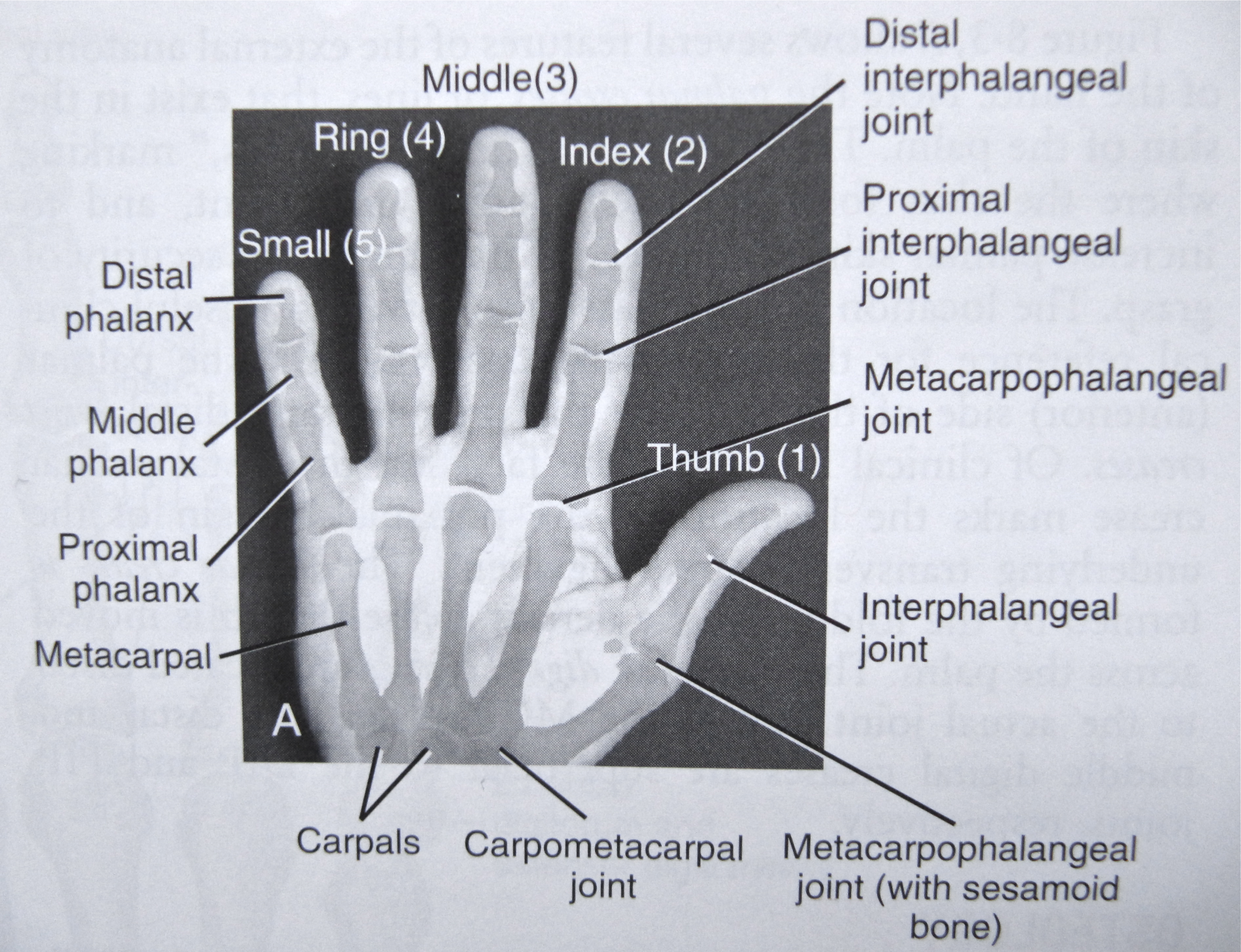
The Hand’s Arches
The hand, like the foot, has three arches: the distal and proximal transverse arches running across the hand, and the longitudinal arch coursing along the hand’s length.
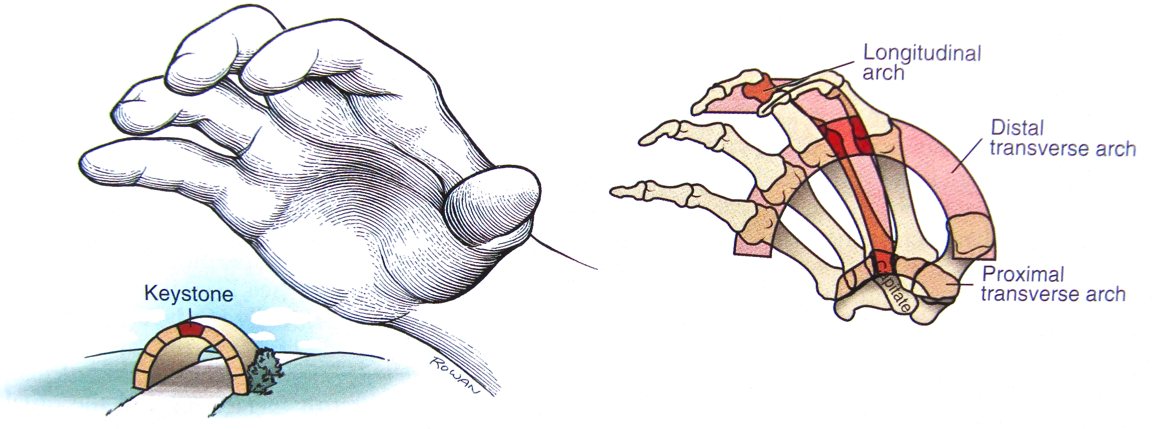
The distal transverse arch lies where the fingers meet the metacarpal bones of the palm. The flexibility of this arch allows the palm to flatten as the hands open and go out in the tor yu with the fingertips up. And with Whip To One Side, it permits the outside first, fourth and fifth metacarpals of the right hand to fold about the more fixed second and third metacarpals while the tips of the fingers come together to form a beak. The two central metacarpals form the keystone of the arch.
The proximal transverse arch arises from the bones of the wrist. Although much more rigid than the distal arch, it opens and flattens over time as a result of placing the hand in Tiger’s Mouth. Gradually, the appearance of the heel of the hand changes.
The longitudinal arch, running the length of the hand, is centered about the second and third metacarpals which run to the index and middle fingers. Stiff at its proximal end where it joins the wrist, this arch is very mobile distally, as active flexion and extension of the fingers demonstrate. Each arch supports the other two, illustrating how elements weave together for strength.
The Soft Tissues of the Hand
Thus far, we’ve referred to the bony architecture of the hand. But layer upon layer of soft tissue contribute to the hand’s structure as well – sheets of fascia, tendons, tendon sheaths, muscles, nerves, arteries, veins and lymphatics.
Without worrying about the names of all these constituents, let’s look quickly at four drawings from Netter that give us a sense of those layers. These are views of the front of the hand; similar layers also exist at the dorsal aspect of the hand.
Don’t get lost in the details. Just note that these successive layers exist beneath the skin of your palm. And because we may not describe this for other regions of the body, remember that this complexity is found everywhere. Which is why we keep things simple and work with metaphors like Tiger’s Mouth when we play at tai chi.
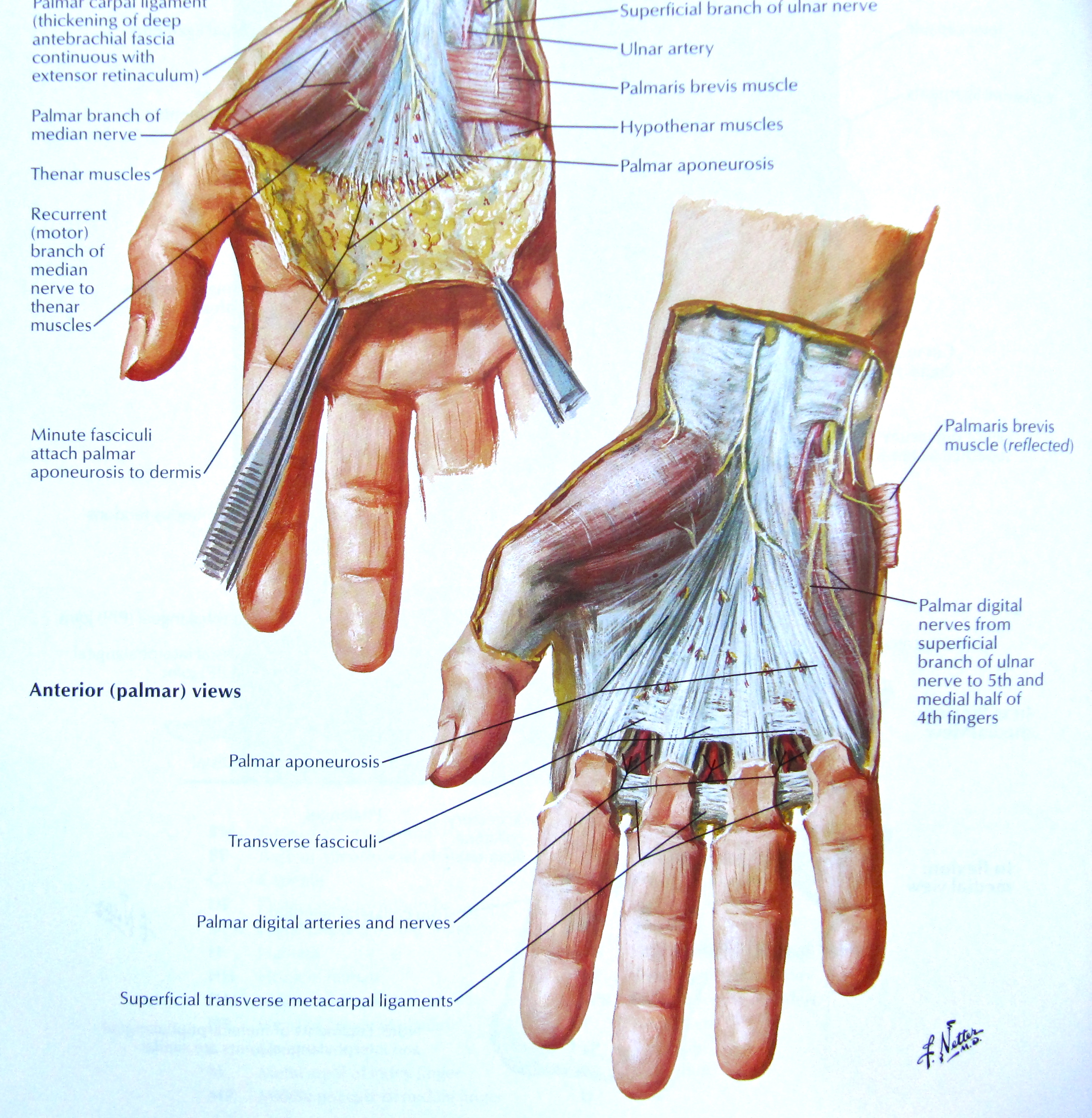
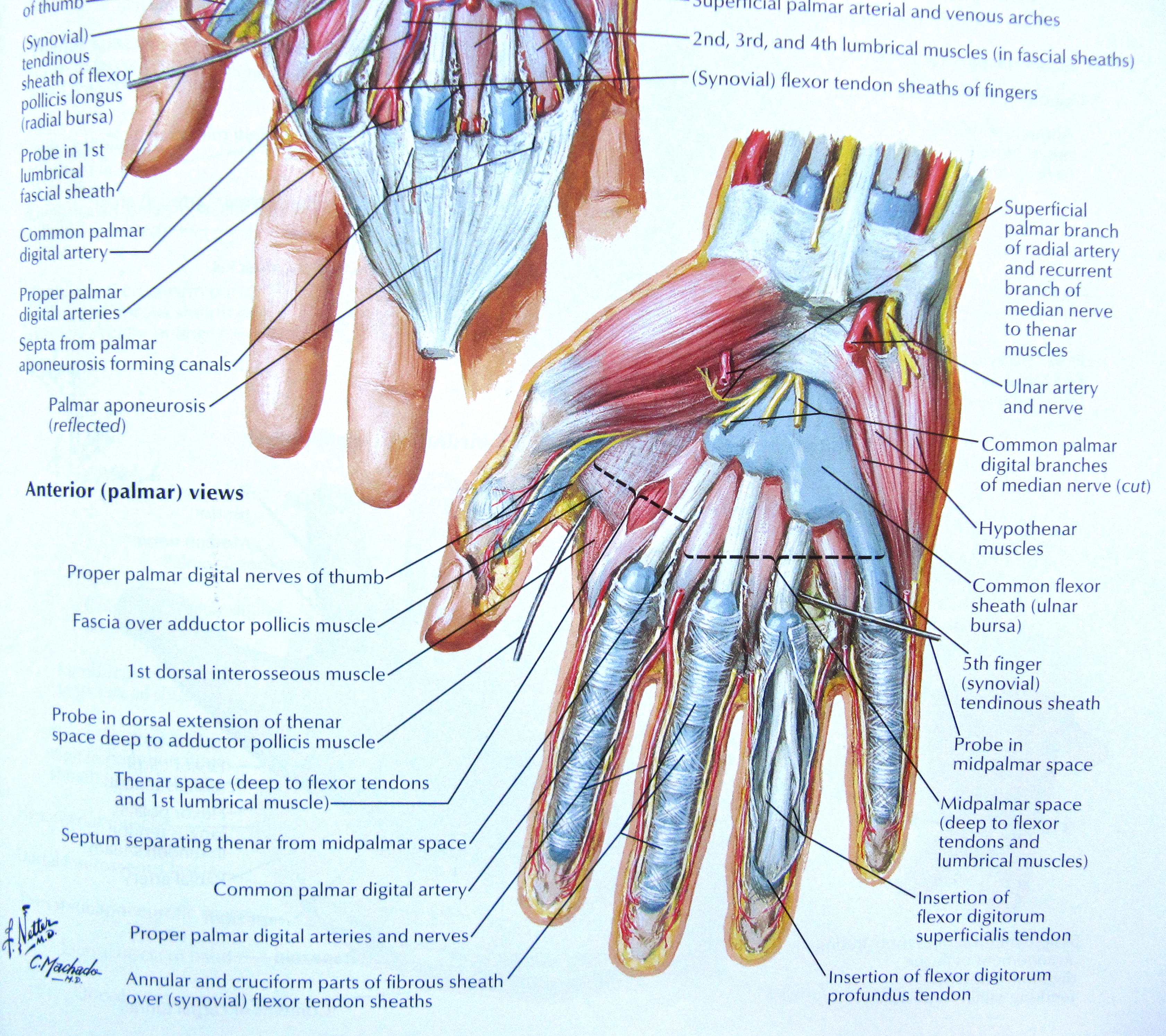
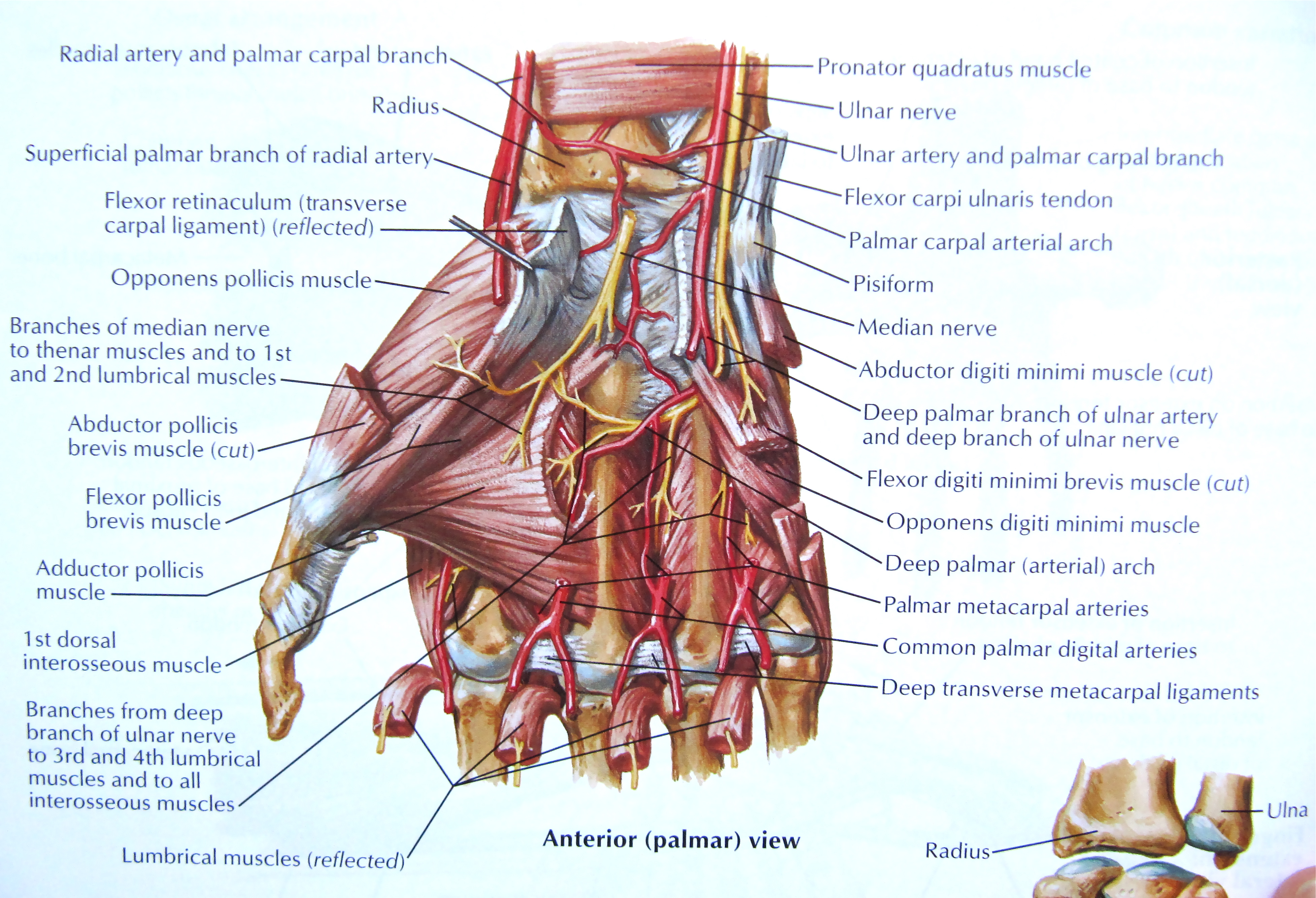
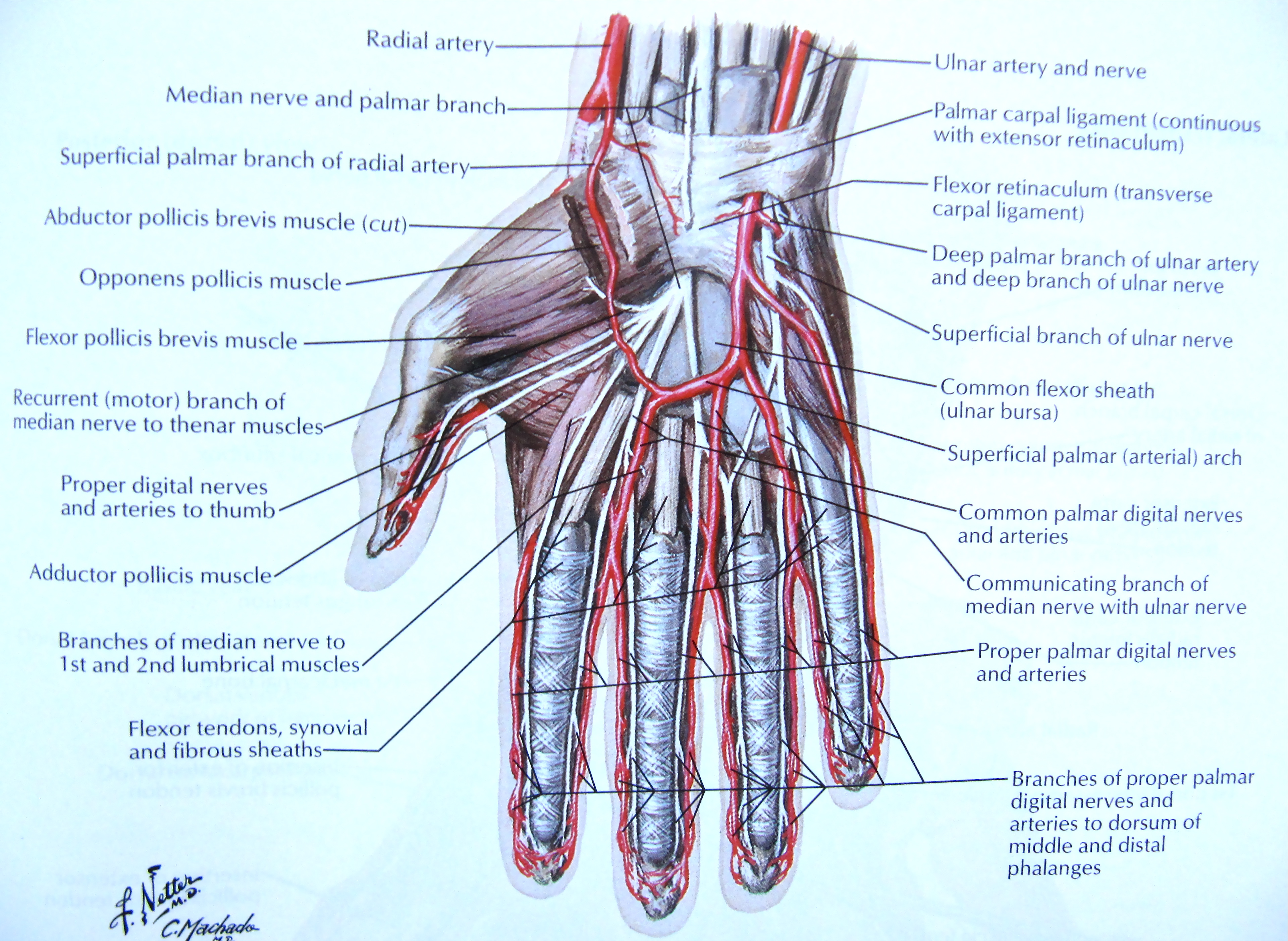
Having spent a few moments reading about anatomy, move one of your hands from a position of rest into Tiger’s Mouth. Feel each of the three arches open up and note the increased sense of structure. Imagine how the Tiger’s Mouth engages all the soft tissues of the palm depicted in the drawings above, elongating them and creating an elastic or return force in the process.
Notice, too, how stretching out all these tissues creates a flow of sensory information into our central nervous system. The hand is an extremely important sensory organ, so important that a large part of the sensory cortex of the brain is dedicated to it. Work done by Dr. Penfield, a Montreal neurosurgeon, gave us images known as sensory and motor homunculi.
A homunculus is a representation of the whole human form, based in this case on the area of cortex devoted to sensing or moving the various body parts.
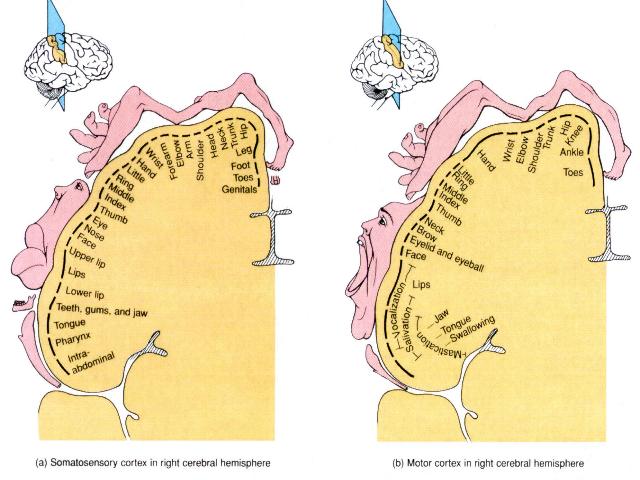
In a very real sense, then, the body has another existence in the brain. The hand, for example, is as much a network of sensory nerves in the brain as it is a physical form.
Another way of expressing the value attributed to sensation coming from the hands is to create an image of what the body would look like if each part grew according to the area of the brain concerned with its sensory perception.

So the hand is a major sensory organ, one that is equally capable of a vast array of intricate movements. When we use our hands with intention in the set, we become more aware of where our hands are and of what they are doing. This enlarges the area of cortex devoted to the sensory data streaming in from the hand. This, in turn, leads us to become even more mindful of the hands. Neuroplasticity in action.
And because the hands are connected to the rest of the body, much like the tip of a whip is linked with its handle, we can use them not only to sense the outer edges of our upper limbs but to increase awareness of the spine, pelvis and feet.
1. Kinesiology of the Musculoskeletal System, Foundations for Rehabilitation, Second Edition, 2010, Donald A. Neumann, Mosby Elsevier, ISBN 978-0-323-03989-5
2. Atlas of Human Anatomy, 4th Edition, Frank H. Netter, 2006, Saunders Elsevier, ISBN 13: 978-1-4160-3385-1

