The Dragon's Head Blog: Notes on Anatomy and Physiology: The Intervertebral Discs
The intervertebral discs play a key role in the life of the healthy spine. Their degeneration is a frequent cause of pain and disability, and a herniated lumbar disc represents the most common reason adults end up with back surgery. And many students first come to the Taoist Tai Chi® arts because of persistent back pain.
So, let’s spend a bit of time reviewing disc structure and behaviour.
As mentioned before, 24 vertebrae stack one on top of the other to connect head to pelvis. Between each pair we find an interposed disc – starting at the junction of the second cervical vertebrae with the third (C2-3) and running all the way down to the joint between the fifth lumbar vertebra and sacrum (L5-S1).
The discs, made of strong but deformable soft tissue, separate the vertebral bodies from one another, thus allowing movement between them.
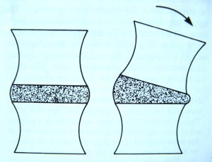
The following cartoon gives you a quick sense of how the discs work as spacers between vertebral bodies.
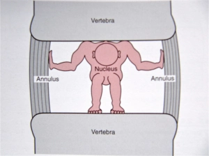
As we’ll see shortly, the discs also serve as shock absorbers and transmit a loading force from one vertebra to the next.
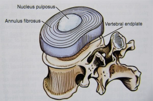
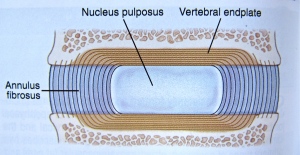
Each disc has 3 components:
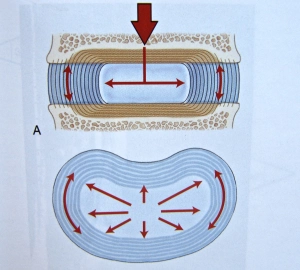
- the nucleus pulposus located towards the centre of the disc. In young people, this material is a gel made mostly of water, giving it the consistency of toothpaste. As a ball of fluid, it deforms with weight-bearing but cannot be compressed. Apply pressure and the nucleus changes shape – without any reduction in its total volume. Pressure exerted on the disc is transmitted radially, much like a compressed water balloon stretches out its walls in all directions.
- the annulus fibrosus: 10-20 densely packed, concentric rings of collagen fiber strands that surround and contain the liquid nucleus like dough encircles the jelly of a doughnut. The collagen fibers arrange themselves in a fixed pattern: their direction alternates in successive rings from left to right, always maintaining an orientation of about 65 degrees off vertical.
- the vertebral endplates. These cartilaginous caps of connective tissue cover most of the top and bottom surfaces of the vertebral bodies. Although tightly bound to the disc by the collagen fibers of the annulus, the endplates are only loosely bound to the vertebral bodies.
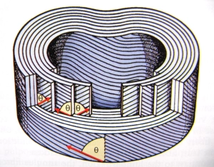
When loaded from above, the height of the nucleus is reduced. It attempts to expand out against the annulus and the collagen rings of the annulus are stretched. The radial pressure exerted by the nucleus is quickly balanced by the elastic tension that develops in the lengthening fibers of the annulus. As a consequence, a 40 kg load produces in a healthy disc only 1 mm of vertical compression and 0.5 mm of radial expansion.
At the same time, the nucleus is constrained in the up-down sense by the endplates and vertebral bodies.
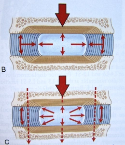
In this way, pressure applied to the nucleus is passed on to both the annulus and the endplates. The fibers of the annulus are braced and prevented from buckling. The disc works as a shock absorber when the spine experiences a rapidly applied force; the force can be momentarily diverted into the annulus, easing the speed with which it must be transmitted down the chain of vertebrae.
Pressure exerted on the endplates transmits part of the load from one vertebra to the next. The facet joints described in the last post also take some of the strain, as do the ligaments, tendons, muscles and thoracolumbar fascia of the back. Compressive forces end up being shared by multiple structures so that no single tissue risks injury. But to work well together this way, all tissues need to stay healthy.
With the removal of a loading force, the elastic energy stored in the collagen rings causes the annulus to recoil and the normal shape of the nucleus is restored.
In the next post, we will look at the pressures to which the discs are typically subjected, some of the changes associated with aging of the spine, and the development of degenerative disc disease.
1. Clinical Anatomy of the Lumbar Spine and Sacrum, Nikolai Bogduk, 1987, Churchill Livingstone, ISBN 0 443 06014 2
2. The Illustrated Guide to Functional Anatomy of the Musculoskeletal System, Rene Cailliet, 2004, AMA press, ISBN 1-57947-408-X
3. Kinesiology of the Musculoskeletal System, Foundations for Rehabilitation, Second Edition, Donald A. Neumann, 2010, Mosby Elsevier, ISBN 978-0-323-03989-5

