The Dragon's Head Blog: Notes on Anatomy and Physiology: Getting the Feel of Tensegrity
We’ve spoken recently of how the body makes use of tensegrity to help hold itself together. We stretch out our soft tissues and they resist further expansion and create a sea of continuous tension that tugs on and supports the bones suspended within it. Soft tissue and bone working together.
Because the soft tissues resist further expansion, they supply an ever-present, wall-to-wall, continuous pull inward on the bones. The bones, by refusing further compression, push outward locally on the soft tissues surrounding them and prevent our structure from collapsing in on itself. We create a form that is less stiff, more resilient than one relying on continuous compression.
Thinking of the body’s architecture this way leads us to view the elastic, distensible elements of the body in a new light.
If we use the word „tendon“ to mean any soft, elastic tissue that contributes to a web of continuous tension, we begin to see many structures of the body as tendon: the lungs that want to spring back once filled; the diaphragm, esophagus, stomach and intestine; the aorta, the great artery that leads freshly oxygenated blood out of the heart to all corners of the body; the veins, lymph channels and nerves, supplied as they are with walls that contain elastic connective tissue; the ligaments and capsule that surround each joint; the muscles and skin and the wrappings of the brain and spinal cord.
All are capable of elastic recoil. All are connected with one another. And all are prone to the ill effects of aging and disuse, and must regularly go through a full range of motion to maintain elasticity and avoid desiccation.
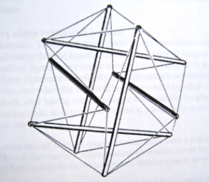
Building a tensegrity structure quickly give us a feel, an intuitive sense, of how the body works in this way. Taking the time to construct an icosahedron is well worth the effort.

What follows is a summary of the instructions for building such a model provided by Thomas Myers in his book, Anatomy Trains, second edition:
- Collect 6 dowels (I used 3/8 inch dowels, each 12 inches long), small screws (12, one for each dowel end) and 24 elastics (Universal Rubber Bands, size 31, 2-1/2 x 1/8 x 1/32 inches, order #00431, worked well for me). When placing the screws in the dowel ends, leave enough shaft visible so that 4 rubber bands may be slipped under the head of each screw. Have another pair of hands available to help with the assembly.
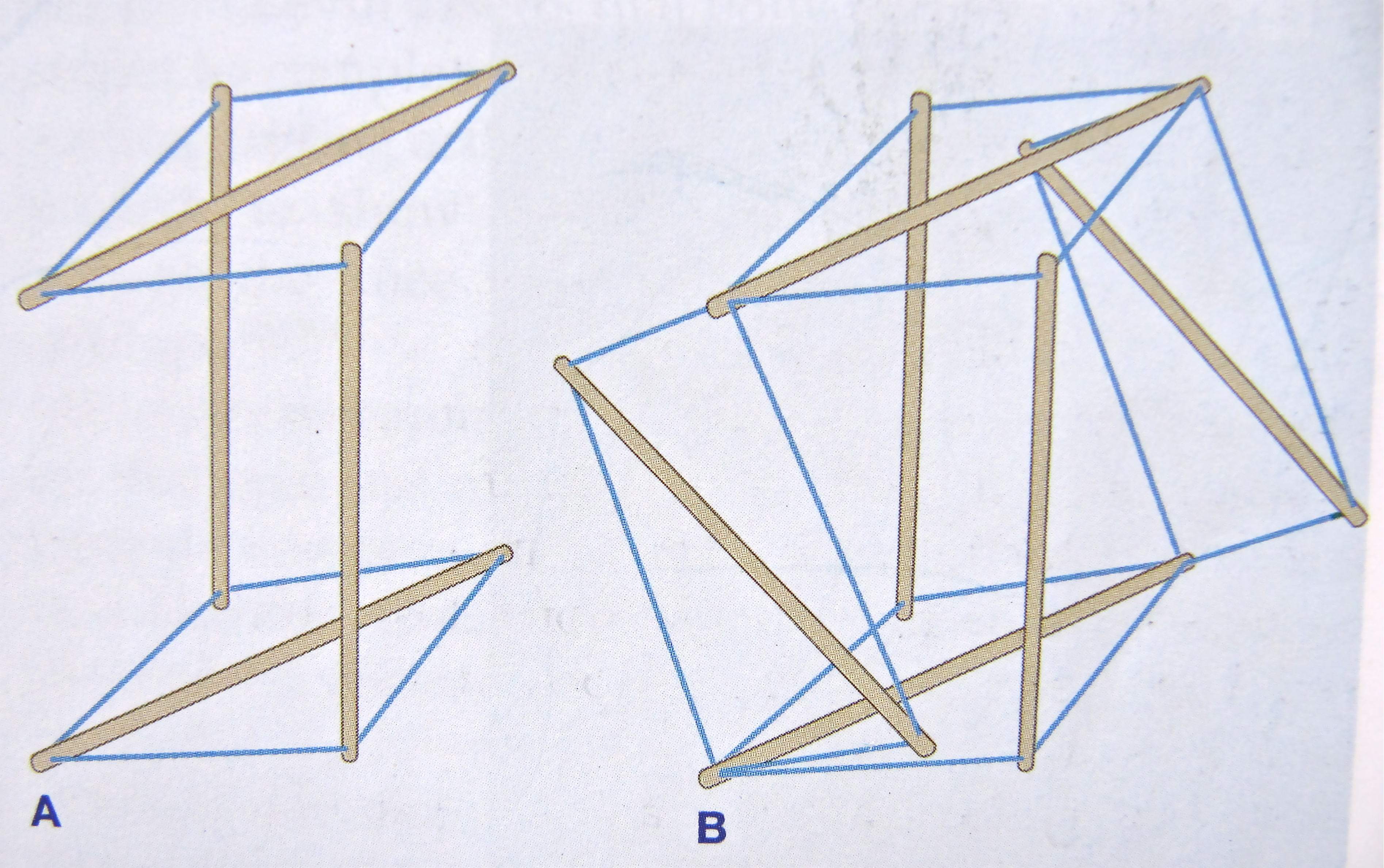
- Take 2 dowels. While holding them vertically and parallel to one another, place a third dowel horizontally between them near the top to form the letter „T“. Connect rubber bands from each of the upper ends of the 2 verticals to each end of the horizontal dowel, using 4 elastics in all.
- Turn the vertical dowels upside down (180 degrees) so that the horizontal dowel now lies on the table. Repeat step 2, attaching 4 elastics from these ends of the uprights to the ends of a new horizontal dowel. You have now formed the capital letter „I“. See fig 2A below.
- Turn the structure 90 degrees so that the 2 horizontal dowels are now upright, creating an „H“ with two crossbars. Make sure those extra set of hands are close by as the next few steps are a bit more difficult. Place a fifth dowel horizontally between the 2 uprights so that this fifth dowel is at right angles to all the others and pointing toward and away from you. Connect the 2 uprights to the ends of the new horizontal dowel. Maintain a sense of humour as elastics fly unexpectedly into the air.
- Turn the structure over and repeat step 4, employing the sixth and last dowel. See figure 2B below.
- Finish by adding the remainder of the elastics, connecting each dowel end to all four adjacent ends, except for the ends of the dowel that is parallel to it. The resulting structure will stand alone, more or less balanced and symmetric, with 3 sets of parallel pairs of dowels. Each dowel end will have 4 elastics running out from it to the other ends nearby while remaining unconnected to its parallel partner. You may need to take an extra turn or two about some of the screws to even out the tension and improve the symmetry.

If the elastics are excessively long, the tendons of the model will be too lax to provide adequate structure. If unduly short, the tight tendons will make it difficult for the structure to alter its shape with changing stresses. Both these situations mirror what happens in the practice hall when our soft tissues are too loose or too tight.
As you play with the model, you appreciate that the rubber bands form a continuous web while the dowels fail to touch one another. Continuous tension, discontinuous compression. Alter a single elastic or dowel and the whole structure responds. Draw two parallel dowels apart and the whole structure expands in all directions – recreating much of the feeling of density, fullness or stickiness that comes with balanced movement and was mentioned in the last note. Push two parallel dowels together and the entire structure contracts.
A complete structure expanding and contracting. Breathing in and breathing out.
Reflections about the body begin to surface:
- does the spine really function as a continuous compression structure? Or do the 24 vertebrae perch precariously one on top of the other, only kept in place by a fabric of soft tissue?
- soft tissue forces contribute greatly to our shape, our posture. By altering those forces, we alter our shape
- Taoist Tai Chi® arts work to create a balance of tension forces within all our soft tissues so that we may move with poise and ease
- tensegrity is one way the body manages potentially injurious loads, distributing, rather than localizing, forces that act on it
- the soft tissues (muscle, tendon, ligament, fascia) are always under some tension. In this way that they contribute to structure. In the cadaver, for instance, sever the anterior and posterior longitudinal ligaments and the ligamentum flavum, leave only the intervertebral discs to hold the vertebral bodies together, and the vertebral column expands
- the angle between elements of an icosahedron is 60 degrees, the same angle taken by the alternating layers of the annulus fibrosus
- Stephen Levin, an orthopedic surgeon, believes that the bony components of the human tensegrity system „kiss“ but are not in compressive opposition with one another. He describes how, during a surgical procedure on a living subject, the articular surfaces of the joints of the knee, ankle, and elbow cannot be forced into contact with one another as long as the surrounding ligaments remain intact. This view is debated by others and runs counter to our usual way of thinking about how the bones stack to transmit weight down through the body. In life, the body likely moves through a continuum between continuous compression and full tensegrity, utilizing different strategies in different circumstances and at different times. What is clear is that our elastic soft tissue, not just our bones, contribute greatly to structure and to our ability to alter shape and move about
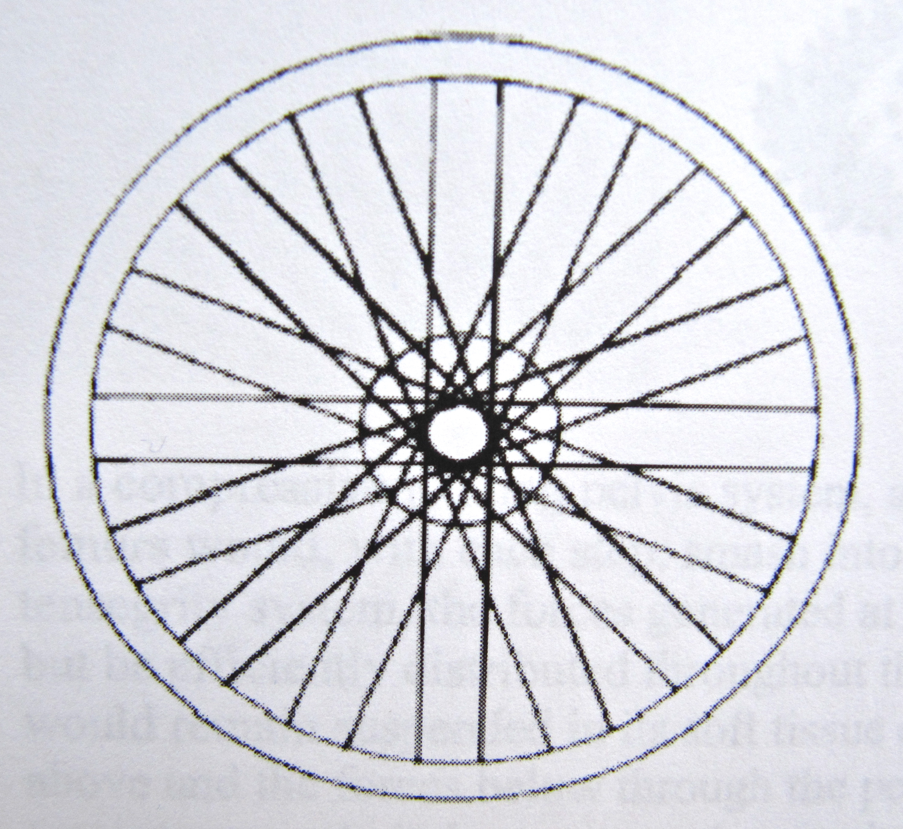
- the shoulder blade doesn’t sit on top of anything. Rather, it hangs off the rib cage, floating in a tension network created by the surrounding muscles and tendons of the shoulder girdle. Much like the hub of a bicycle wheel is suspended in a tension network of spokes.


1. Anatomy Trains, Second Edition, 2009, Thomas W. Myers, Churchill Livingston Elsevier, ISBN: 978-0-443-10283-7
2. Dr. Stephen M. Levin, orthopedic surgeon

